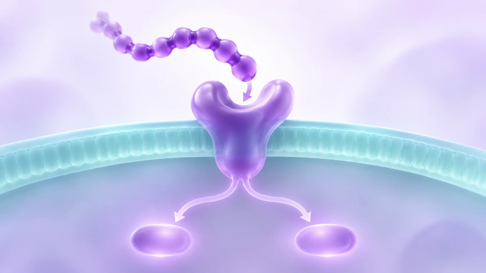
Why does the receptor have three names?
Read the kisspeptin papers from 2001 in a row and a small mystery appears. One talks about GPR54, another about AXOR12 and a third about hOT7T175. It looks at first as though kisspeptin turned up at three receptors. The nomenclature report from the International Union of Basic and Clinical Pharmacology brings those older labels together under the kisspeptin receptor.
At the time, researchers called it an orphan receptor. They could identify the receptor, but its naturally occurring partner was still unknown. The discovery papers had to join up several kinds of evidence. A signal in a cell could point to a promising molecule. Someone still had to identify what the molecule was, show how it interacted with the receptor and find out whether the interaction changed a cellular readout.
From a tissue sample to an identified peptide
Kotani and colleagues isolated three peptides from human placental tissue. They were 54, 14 and 13 amino acids long, and shared the same amidated ending. The authors traced them to the product of the KISS1 gene and called them kisspeptins. Ohtaki and colleagues, working separately, reported a 54-residue amidated peptide from the same type of tissue and named it metastin. Ohtaki’s paper lists a Takeda research affiliation.
This part of the discovery answers where the researchers found the molecule and what they identified. Keep the tissue sample separate from the cell model in your reading notes. The placental preparation was the source of the peptides. The receptor experiments used cultured cells to test whether the isolated molecules had the activity the teams were looking for. A paper can contain both kinds of evidence even when they appear in the same paragraph of the abstract.
Sources: Kotani and colleagues, Journal of Biological Chemistry (2001): The metastasis suppressor gene KiSS-1 encodes kisspeptins, the natural ligands of the orphan G protein-coupled receptor GPR54 · Ohtaki and colleagues, Nature (2001): Metastasis suppressor gene KiSS-1 encodes peptide ligand of a G-protein-coupled receptor
What made the receptor assignment convincing?
Kotani’s group examined binding to human and rat GPR54 expressed in CHO-K1 cells, a cell line derived from Chinese hamster ovary. They also recorded cellular responses, including a change in intracellular calcium. The binding result put the peptide and receptor in the same experimental interaction. The calcium signal showed a response downstream of that interaction in the cell preparation.
This is where it helps to read the figure titles before the authors’ conclusion. If the experiment reports that a candidate displaces a labelled ligand, the evidence concerns binding under those conditions. If the experiment reports calcium moving inside a cell, the evidence concerns a cellular response. Look for the comparison that connects each observation to the receptor. A bright signal by itself could have several explanations, especially in a cell with its own native signalling machinery.
Also notice which species each label describes. A human receptor expressed in a hamster-derived cell line is a way to study that receptor in a specified cell system. The species of the receptor and the origin of the host cell are two different details to retain when comparing results.
A second starting point led to the same pairing
Muir and colleagues began from the receptor side. They cloned the human receptor they called AXOR12, expressed it in mammalian cells and used the responses to investigate candidate agonists. The study reported strong responses to peptides derived from KISS1. The publication lists a GlaxoSmithKline discovery biology affiliation for the first author.
Compare the questions the teams brought to the problem. The tissue isolation work connected an observed activity to molecules recovered from a biological source. The receptor-focused screen could flag candidates in a cell model and test how strongly the assay responded to peptides derived from the gene product. The later nomenclature report groups the three discovery papers together because their results converge on the same pairing. Each experiment still has its own sample and readout.
Sources: Muir and colleagues, Journal of Biological Chemistry (2001): AXOR12, a novel human G protein-coupled receptor, activated by the peptide KiSS-1 · Kirby and colleagues, Pharmacological Reviews (2010): International Union of Basic and Clinical Pharmacology. LXXVII. Kisspeptin receptor nomenclature, distribution, and function
Finding receptor RNA opens a different question
Muir’s team also measured receptor RNA across tissue samples. That tells the reader where the study detected a transcript for the receptor. The activity experiment has a different subject. It records how the cells used in the assay respond when they encounter a candidate peptide. To connect the two observations, a researcher would still need to establish which cells in the tissue make receptor protein and how those cells respond in their own setting.
When reading a discovery paper, mark each panel as evidence of identity, binding, response or distribution. A tissue with abundant transcript may be a good place to ask a new question about the receptor. The abundance itself cannot answer that question.
What the number in KP-10 tells you
The numbers in KP-54 and KP-10 give the number of residues. KP-10 shares the final ten residues of the longer peptide and has been shown to bind and activate the receptor in vitro. That makes a shorter fragment useful for asking which part of the sequence can carry activity in an assay.
When you encounter a result reported simply for kisspeptin, look for the fragment name in the methods or figure caption. Record the species of the receptor and the readout alongside it. Even if two fragments both give a response, a comparison of their behaviour depends on which response was measured. The exact material in the paper also needs its own entry when you compare it with separate analytical documentation for a supplied sample.
Frequently asked questions
Is KISS1 the receptor gene?
KISS1 encodes the precursor from which kisspeptins are derived. KISS1R names the gene for the kisspeptin receptor. Older reports may use GPR54, AXOR12 or hOT7T175 when discussing the receptor.
What does metastin refer to in the 2001 paper?
Ohtaki’s group used metastin for the 54-residue peptide it recovered from human placenta. The later nomenclature report calls this peptide kisspeptin-54, or KP-54.
Sources: Ohtaki and colleagues, Nature (2001): Metastasis suppressor gene KiSS-1 encodes peptide ligand of a G-protein-coupled receptor · Kirby and colleagues, Pharmacological Reviews (2010): International Union of Basic and Clinical Pharmacology. LXXVII. Kisspeptin receptor nomenclature, distribution, and function
Was the calcium signal a measurement of how much peptide was bound?
It measured a cellular response. For evidence about association with the receptor, look at the binding results. Kotani’s group reported both types of measurement in receptor-expressing cell preparations.
Sources
- The metastasis suppressor gene KiSS-1 encodes kisspeptins, the natural ligands of the orphan G protein-coupled receptor GPR54Kotani and colleagues, Journal of Biological Chemistry · 2001 · DOI: 10.1074/jbc.M104847200
- Metastasis suppressor gene KiSS-1 encodes peptide ligand of a G-protein-coupled receptorOhtaki and colleagues, Nature · 2001 · DOI: 10.1038/35079135
- AXOR12, a novel human G protein-coupled receptor, activated by the peptide KiSS-1Muir and colleagues, Journal of Biological Chemistry · 2001 · DOI: 10.1074/jbc.M102743200
- International Union of Basic and Clinical Pharmacology. LXXVII. Kisspeptin receptor nomenclature, distribution, and functionKirby and colleagues, Pharmacological Reviews · 2010 · DOI: 10.1124/pr.110.002774
Published by EliteGen Research for research education. Studies describe their own materials and experimental conditions; they do not establish the identity, quality, or suitability of an EliteGen product. This article does not provide guidance for personal, human, or veterinary use.