One paper, two things to follow
Read a paper about a small peptide and you can find several charts on the same page. One tracks material associated with a cell. Another records a change in light. A third looks at protein on a blot. Taken together they tell a story, but every panel gets there by measuring something different.
The KPV study is a good place to practice reading across those panels. Start with a line down the middle of your notes. On one side, put observations about where the peptide goes. On the other, put changes the cells show after exposure. The space between the two columns is where the study’s argument has to do its work.
Follow the tracer through Figure 4
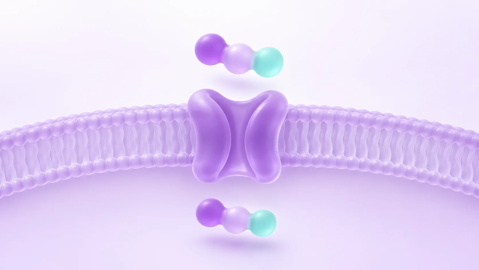
KPV is a three-residue peptide made up of lysine, proline and valine. Dalmasso and colleagues investigated its relationship with PepT1, a membrane protein that transports small peptides. Their 2008 paper includes experiments with human-derived intestinal epithelial cell lines and Jurkat T cells, along with separate chemically induced mouse colitis models.
Figure 4 begins with a competition experiment in Caco2-BBE cells. KPV reduced uptake of a labelled reference substrate. A later panel follows radiolabelled KPV itself. The first comparison asks whether KPV changes the reference substrate’s uptake. The second gives the researchers a way to track KPV uptake more directly.
Why do both panels matter? If you watched only the reference substrate, you would be reading what happened to the reference. A decline in that signal can suggest an interaction, while tracking the labelled test peptide addresses the more specific question of whether that peptide enters the cells.
Put the next PepT1 paper alongside it
In 2010, Dalmasso and colleagues examined Tri-DAP, a bacterial cell-wall fragment. Their epithelial-cell results linked PepT1 to uptake and an increase in inflammatory signalling. When they reduced expression of PepT1 in one cell model, components of the response were delayed or reduced. Expression of PepT1 in a second model strengthened the response relative to the comparison cells.
Read that result alongside the 2008 KPV paper, where several measures of inflammatory signalling went down under the tested conditions. A transport route can help explain access to the cell. The chemical material, the cell’s state and the question asked of the cell still matter to the response.
This is a comparison between studies. It works best as a check on your reading of the word transporter. The papers used different materials, and they were not designed to rank those materials against one another.
Sources: Dalmasso and colleagues, American Journal of Physiology—Gastrointestinal and Liver Physiology (2010): PepT1 mediates transport of the proinflammatory bacterial tripeptide L-Ala-{gamma}-D-Glu-meso-DAP in intestinal epithelial cells · Dalmasso and colleagues, Gastroenterology (2008): PepT1-mediated tripeptide KPV uptake reduces intestinal inflammation
What the light in Figure 1 is reporting
The 2008 paper used a luciferase reporter to follow NF-κB signalling. Figure 1 also includes measurements of signalling proteins and IL-8 RNA and protein. A pattern across different kinds of readout gives readers more to work with than a single light signal.
Luciferase makes light that the instrument can count. In a reporter experiment, the light stands in for the process the researchers want to follow. Auld and Inglese explain in the Assay Guidance Manual that compounds can sometimes change the detection system itself. That is a general issue with reporter assays, and it gives you a reason to check which other measurements a study actually included.
An independent readout addresses the biology by another route. A counterscreen probes a particular source of error, such as interference with detection. These are distinct ways of testing an interpretation. When you find either in a paper, ask which conclusion it helps you trust and which questions it leaves open.
Sources: Dalmasso and colleagues, Gastroenterology (2008): PepT1-mediated tripeptide KPV uptake reduces intestinal inflammation · Auld and Inglese, Assay Guidance Manual, NCBI Bookshelf (2018): Interferences with Luciferase Reporter Enzymes
What would a missing control leave you wondering?
Suppose a fictional paper included two measurements: it detected a peptide in a cell sample, and it recorded a change in a signalling pathway in a separate experiment. Those facts could sit side by side even if the pathway changed for a different reason. The comparison you would look for next is one that connects the two observations by asking whether the response changes when the route is unavailable.
Keep the question small enough to answer. A change in response associated with a transporter may tell you something about how material got to the relevant part of the system. A claim about the exact molecule it contacts once there adds another step. If a figure legend makes that step sound automatic, the methods are a better place to check.
Put the model next to the claim
Try giving each panel a caption of your own. “In this cell line, this tracer changed.” “In this stimulated-cell comparison, this signalling readout changed.” Both sentences can be true without settling what KPV contacts after entry. To answer that question, you would need evidence that pinpoints the molecular interaction itself.
Use the same habit when the paper moves from cultured cells to mice. A human-derived cell line gives the researchers a laboratory model. The mouse work supplies observations from a separate model. Neither model measures an outcome in a person.
The 2008 paper acknowledges NIH and foundation support. Both Dalmasso papers declare no conflicts of interest. Those disclosures belong beside the study details when you keep a record of the evidence.
Sources: Dalmasso and colleagues, Gastroenterology (2008): PepT1-mediated tripeptide KPV uptake reduces intestinal inflammation · Dalmasso and colleagues, American Journal of Physiology—Gastrointestinal and Liver Physiology (2010): PepT1 mediates transport of the proinflammatory bacterial tripeptide L-Ala-{gamma}-D-Glu-meso-DAP in intestinal epithelial cells
Frequently asked questions
Why did the researchers measure a reference substrate as well as KPV itself?
The reference helps show that KPV changes uptake of material normally carried by PepT1. Following labelled KPV addresses what happens to KPV itself. Together the panels connect the comparison to the test peptide.
Does finding uptake also identify an intracellular binding target?
Uptake evidence can support a route of entry. A signalling change measured afterward can narrow the questions worth asking, but the identity of a direct target would require a separate line of evidence.
Do the two Dalmasso papers disagree about PepT1?
They examine different molecules. The contrast is in the downstream responses they report. Both studies give transport evidence in their particular models, and neither design makes a side-by-side comparison of KPV and Tri-DAP.
Sources: Dalmasso and colleagues, Gastroenterology (2008): PepT1-mediated tripeptide KPV uptake reduces intestinal inflammation · Dalmasso and colleagues, American Journal of Physiology—Gastrointestinal and Liver Physiology (2010): PepT1 mediates transport of the proinflammatory bacterial tripeptide L-Ala-{gamma}-D-Glu-meso-DAP in intestinal epithelial cells
What does a luciferase reporter measure directly?
The direct measurement is light. Researchers connect it to the pathway through the design of the reporter and the controls, then use additional readouts to test that interpretation.
Sources
- PepT1-mediated tripeptide KPV uptake reduces intestinal inflammationDalmasso and colleagues, Gastroenterology · 2008 · DOI: 10.1053/j.gastro.2007.10.026
- PepT1 mediates transport of the proinflammatory bacterial tripeptide L-Ala-{gamma}-D-Glu-meso-DAP in intestinal epithelial cellsDalmasso and colleagues, American Journal of Physiology—Gastrointestinal and Liver Physiology · 2010 · DOI: 10.1152/ajpgi.00527.2009
- Interferences with Luciferase Reporter EnzymesAuld and Inglese, Assay Guidance Manual, NCBI Bookshelf · 2018
Published by EliteGen Research for research education. Studies describe their own materials and experimental conditions; they do not establish the identity, quality, or suitability of an EliteGen product. This article does not provide guidance for personal, human, or veterinary use.