Start with the chain
A peptide can be short enough to write on one line. The experiments built around it can still fill a journal issue. That is part of the appeal: a scientist can name a small molecule precisely and then ask what it does in a system that has many moving parts.
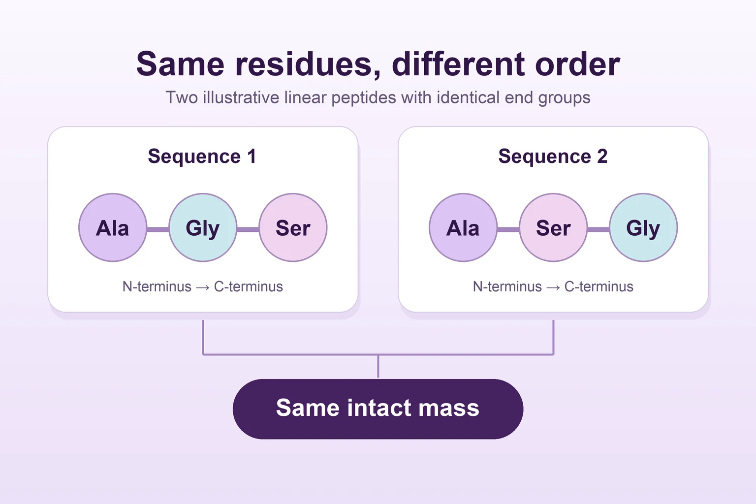
The chain consists of amino acids connected by peptide bonds. Amino acids are the same class of building blocks that make up proteins. The word peptide commonly refers to a shorter chain, but the label is only the beginning of the description. A useful starting point is the material's sequence, the order in which those amino acids are joined. That gives you something much more specific to take into a paper than the name on the first page.
Sources: National Human Genome Research Institute, NIH: Peptide
The same ingredients can make different chains
Imagine two cards that list the same four amino acids in a different order. A tally of the ingredients would look the same. The order of the chain would differ, and the chemical groups would sit in different positions along it. That is why a sequence is more informative than a list of what went into a molecule.
The sequence gives the reader a way to ask sharper questions about structure. Where are the charged groups? Could part of the chain contact another molecule? Does the experiment concern a single chain or something assembled from many chains? The protein-structure chapter in Molecular Biology of the Cell describes the connection between sequence, chemical interactions and three-dimensional shape. For a short peptide, the surrounding solution or a binding partner can also matter to the shapes it samples. Read the sequence as a starting point for investigating structure.
When a peptide meets a receptor
Some peptides serve as signals. A receptor recognizes the signal and can set off changes inside the cell. The interesting part for a reader is how an experiment takes that broad idea apart. One assay might ask whether the peptide and receptor interact. A second might look for a change in an intracellular messenger. A third could observe where the receptor goes after the signal arrives.
You can read all three figures with the same question in mind: what did the instrument actually detect? For a binding figure, the readout might reflect occupancy or competition with a reference ligand. A signaling figure measures a step downstream. The cells provide the surrounding machinery for that step, so two models can give different results even when the peptide has the same name in both figure legends.
The day a short chain made a visible material
Receptor signaling is only one corner of peptide science. In 1993, Zhang and colleagues reported that a 16-residue peptide assembled into a membrane under their experimental conditions. The scanning electron microscopy images showed interwoven filaments. The researchers were studying what many copies of a small chain could make together.
Notice how that example changes the meaning of the word structure. A sequence tells you about the arrangement of amino acids within one molecule. A microscopy image of an assembled material describes a much larger object. In this paper, the membrane and its filaments are part of the result. A receptor assay would give a completely different window onto the same broad family of molecules.
Read the instrument output before naming the effect
A paper might say it measured viable cells. What you see in the figure, though, could be a light signal or a color change. The Assay Guidance Manual explains how viability assays use markers associated with living cells. Each assay has a measurement chain: there is the biological property the researchers care about, the marker they use and the signal the reader sees.
That distinction gives you a way to diagnose a surprisingly strong or weak result. Ask whether the study included a control for the detection step. A compound that affects the chemistry used to generate a signal can change the reading before you have an explanation for what happened to the cells. A separate measurement can help distinguish those possibilities. The methods section is where that reasoning becomes visible.
Sources: Riss and colleagues, Assay Guidance Manual, NCBI Bookshelf: Cell Viability Assays
A small exercise for your next paper
Pick one figure and describe it without using the paper's headline. Write one sentence that names the material and the model. Write a second that describes the instrument output and the comparison. Then ask what conclusion you would feel comfortable drawing from those two sentences alone.
This exercise works for a receptor plot, a chromatogram and a microscopy image. It also helps you spot a gap between a published study and a material specification. The paper can tell you which material the scientists studied. A certificate or test report answers questions about the particular sample it describes. Keep both documents in view when identity matters.
One more detail is worth saving: what the paper called its comparison. A solvent-only control, a related peptide and a known reference ligand each answer a different question. If you are reading two papers side by side, write the comparison beside the measurement before deciding whether the results agree.
Frequently asked questions
Is there a single length where a peptide becomes a protein?
Shorter chains are commonly called peptides, and proteins are built from polypeptide chains. Usage varies. The exact sequence and the molecular form used in the experiment give you a firmer basis for comparing papers.
Sources: National Human Genome Research Institute, NIH: Peptide · Alberts and colleagues, Molecular Biology of the Cell, fourth edition (2002): The Shape and Structure of Proteins
Can two peptides have the same amino acids in a different order?
Yes. A composition lists the ingredients, while a sequence tells you how they are ordered. Changing the order changes which chemical groups sit next to one another along the chain.
Does every peptide act as a signal?
Peptide describes the chemistry of a chain. Some peptides are studied as signals, while the 1993 membrane example concerns how many molecules assemble into a material. The experiment tells you which question the researchers were asking.
Sources: Alberts and colleagues, Molecular Biology of the Cell, fourth edition (2002): General Principles of Cell Communication · Zhang and colleagues, PNAS (1993): Spontaneous assembly of a self-complementary oligopeptide to form a stable macroscopic membrane
Where should a new reader start in a study?
Choose one figure. Its legend and methods usually tell you what the model was, what the instrument measured and which comparison gives the result its meaning.
Sources
- PeptideNational Human Genome Research Institute, NIH
- The Shape and Structure of ProteinsAlberts and colleagues, Molecular Biology of the Cell, fourth edition · 2002
- General Principles of Cell CommunicationAlberts and colleagues, Molecular Biology of the Cell, fourth edition · 2002
- Spontaneous assembly of a self-complementary oligopeptide to form a stable macroscopic membraneZhang and colleagues, PNAS · 1993 · DOI: 10.1073/pnas.90.8.3334
- Cell Viability AssaysRiss and colleagues, Assay Guidance Manual, NCBI Bookshelf
Published by EliteGen Research for research education. Studies describe their own materials and experimental conditions; they do not establish the identity, quality, or suitability of an EliteGen product. This article does not provide guidance for personal, human, or veterinary use.